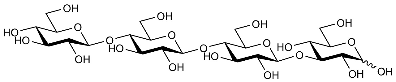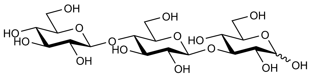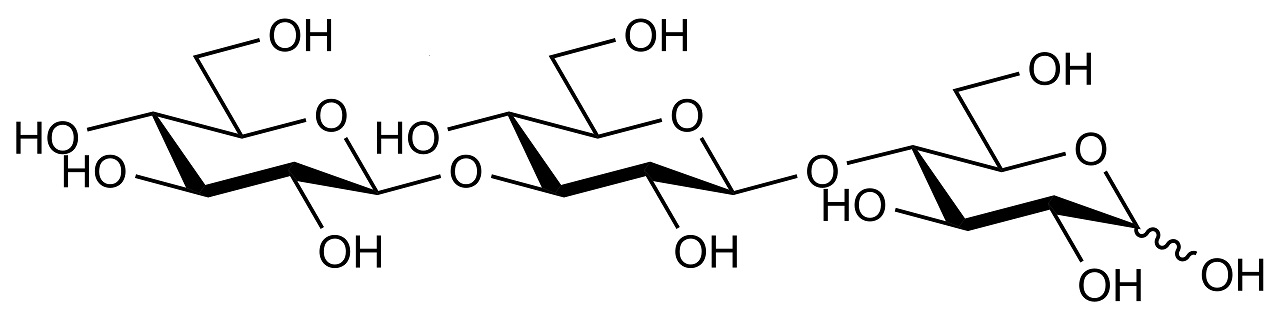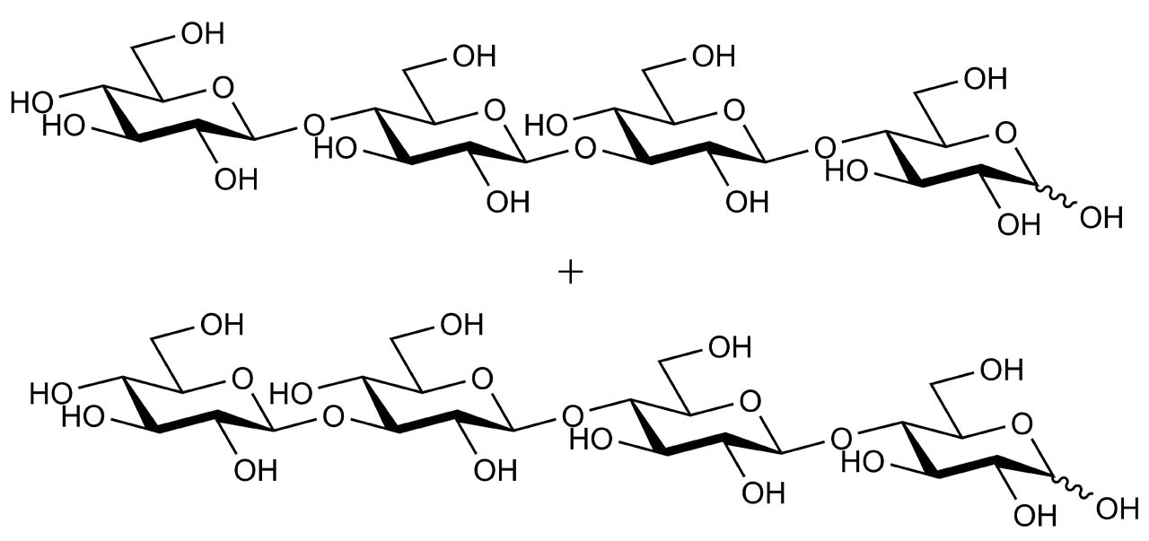
32-β-D-Cellobiosyl-cellobiose +
33-β-D-Glucosyl-cellotriose
30 mg
This product has been discontinued
58484-04-1
This product has been discontinued (read more).
High purity 32-β-D-Cellobiosyl-cellobiose + 33-β-D-Glucosyl-cellotriose mixture for use in research, biochemical enzyme assays and in vitro diagnostic analysis.
These tetrasaccharides are produced on hydrolysis of 1:3,1:4-β-D-glucan by cellulase.
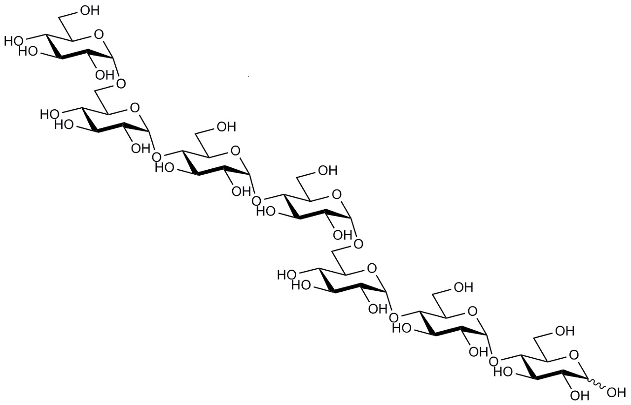
See other products on our high purity oligosaccharides product list.
Related Products
Similar Products
Associated Enzymes
Related Products
Publications

Versatile high resolution oligosaccharide microarrays for plant glycobiology and cell wall research.
Pedersen, H. L., Fangel, J. U., McCleary, B., Ruzanski, C., Rydahl, M. G., Ralet, M. C., Farkas, V., Von Schantz, L., Marcus, S. E., Andersen, M.C. F., Field, R., Ohlin, M., Knox, J. P., Clausen, M. H. & Willats, W. G. T. (2012). Journal of Biological Chemistry, 287(47), 39429-39438.
Versatile high resolution oligosaccharide microarrays for plant glycobiology and cell wall research.
Pedersen, H. L., Fangel, J. U., McCleary, B., Ruzanski, C., Rydahl, M. G., Ralet, M. C., Farkas, V., Von Schantz, L., Marcus, S. E., Andersen, M.C. F., Field, R., Ohlin, M., Knox, J. P., Clausen, M. H. & Willats, W. G. T. (2012). Journal of Biological Chemistry, 287(47), 39429-39438.
Microarrays are powerful tools for high throughput analysis, and hundreds or thousands of molecular interactions can be assessed simultaneously using very small amounts of analytes. Nucleotide microarrays are well established in plant research, but carbohydrate microarrays are much less established, and one reason for this is a lack of suitable glycans with which to populate arrays. Polysaccharide microarrays are relatively easy to produce because of the ease of immobilizing large polymers noncovalently onto a variety of microarray surfaces, but they lack analytical resolution because polysaccharides often contain multiple distinct carbohydrate substructures. Microarrays of defined oligosaccharides potentially overcome this problem but are harder to produce because oligosaccharides usually require coupling prior to immobilization. We have assembled a library of well characterized plant oligosaccharides produced either by partial hydrolysis from polysaccharides or by de novo chemical synthesis. Once coupled to protein, these neoglycoconjugates are versatile reagents that can be printed as microarrays onto a variety of slide types and membranes. We show that these microarrays are suitable for the high throughput characterization of the recognition capabilities of monoclonal antibodies, carbohydrate-binding modules, and other oligosaccharide-binding proteins of biological significance and also that they have potential for the characterization of carbohydrate-active enzymes.

Degradative GH5 β-1, 3-1, 4-glucanase PpBglu5A for glucan in Paenibacillus polymyxa KF-1.
Yuan, Y., Zhang, X., Zhang, H., Wang, W., Zhao, X., Gao, J. & Zhou, Y. (2020). Process Biochemistry, 98, 183-192.
Degradative GH5 β-1, 3-1, 4-glucanase PpBglu5A for glucan in Paenibacillus polymyxa KF-1.
Yuan, Y., Zhang, X., Zhang, H., Wang, W., Zhao, X., Gao, J. & Zhou, Y. (2020). Process Biochemistry, 98, 183-192.
A novel β-1,3-1,4-glucanase in the glycoside hydrolase family 5 (GH5) has been identified in the secretome of Paenibacillus polymyxa KF-1. The recombinant GH5 enzyme PpBglu5A shows broad substrate specificity, with strong lichenase activity, medium β-1,3-glucanase activity, and minimal cellulase activity. Barley β-glucan, lichenan, curdlan, and carboxymethyl cellulose are hydrolyzed to varying degrees by PpBglu5A, with the highest catalytic activity being observed with barley β-glucan. Hydrolysates from barley β-glucan or lichenan are primarily glucan oligosaccharides with degrees of polymerization from 2 to 4. PpBglu5A also hydrolyzes oat bran into oligosaccharides mainly consisted of di-, tri-, and tetra- oligosaccharides that are useful in the preparation of gluco-oligosaccharides. In addition to hydrolytic activity, transglycosylation was also observed with PpBglu5A and cellotriose as substrate. An in vitro assay indicated that the recombinant PpBglu5A has antifungal activity and can inhibit the growth of Canidia albicans. These results suggest that PpBglu5A exhibits unique properties and may be useful as an antifungal agent.
Poaceae-specific cell wall-derived oligosaccharides activate plant immunity via OsCERK1 during Magnaporthe oryzae infection in rice.
Yang, C., Liu, R., Pang, J., Ren, B., Zhou, H., Wang, G., wang, E. & Liu, J. (2021). Nature Communications, 12(1), 1-13.
Poaceae-specific cell wall-derived oligosaccharides activate plant immunity via OsCERK1 during Magnaporthe oryzae infection in rice.
Yang, C., Liu, R., Pang, J., Ren, B., Zhou, H., Wang, G., wang, E. & Liu, J. (2021). Nature Communications, 12(1), 1-13.
Many phytopathogens secrete cell wall degradation enzymes (CWDEs) to damage host cells and facilitate colonization. As the major components of the plant cell wall, cellulose and hemicellulose are the targets of CWDEs. Damaged plant cells often release damage-associated molecular patterns (DAMPs) to trigger plant immune responses. Here, we establish that the fungal pathogen Magnaporthe oryzae secretes the endoglucanases MoCel12A and MoCel12B during infection of rice (Oryza sativa). These endoglucanases target hemicellulose of the rice cell wall and release two specific oligosaccharides, namely the trisaccharide 31-β-D-Cellobiosyl-glucose and the tetrasaccharide 31-β-D-Cellotriosyl-glucose. 31-β-D-Cellobiosyl-glucose and 31-β-D-Cellotriosyl-glucose bind the immune receptor OsCERK1 but not the chitin binding protein OsCEBiP. However, they induce the dimerization of OsCERK1 and OsCEBiP. In addition, these Poaceae cell wall-specific oligosaccharides trigger a burst of reactive oxygen species (ROS) that is largely compromised in oscerk1 and oscebip mutants. We conclude that 31-β-D-Cellobiosyl-glucose and 31-β-D-Cellotriosyl-glucose are specific DAMPs released from the hemicellulose of rice cell wall, which are perceived by an OsCERK1 and OsCEBiP immune complex during M. oryzae infection in rice.
FGB1 and WSC3 are in planta‐induced β‐glucan‐binding fungal lectins with different functions.
Wawra, S., Fesel, P., Widmer, H., Neumann, U., Lahrmann, U., Becker, S., Hehemann, J. H., Langen, G. & & Zuccaro, A. (2019). New Phytologist, 222(3), 1493-1506.
FGB1 and WSC3 are in planta‐induced β‐glucan‐binding fungal lectins with different functions.
Wawra, S., Fesel, P., Widmer, H., Neumann, U., Lahrmann, U., Becker, S., Hehemann, J. H., Langen, G. & & Zuccaro, A. (2019). New Phytologist, 222(3), 1493-1506.
In the root endophyte Serendipita indica, several lectin‐like members of the expanded multigene family of WSC proteins are transcriptionally induced in planta and are potentially involved in β-glucan remodeling at the fungal cell wall. Using biochemical and cytological approaches we show that one of these lectins, SiWSC3 with three WSC domains, is an integral fungal cell wall component that binds to long‐chain β1‐3‐glucan but has no affinity for shorter β1‐3‐ or β1‐6‐linked glucose oligomers. Comparative analysis with the previously identified β-glucan‐binding lectin SiFGB1 demonstrated that whereas SiWSC3 does not require β1‐6‐linked glucose for efficient binding to branched β1‐3‐glucan, SiFGB1 does. In contrast to SiFGB1, the multivalent SiWSC3 lectin can efficiently agglutinate fungal cells and is additionally induced during fungus-fungus confrontation, suggesting different functions for these two β-glucan‐binding lectins. Our results highlight the importance of the β-glucan cell wall component in plant–fungus interactions and the potential of β-glucan‐binding lectins as specific detection tools for fungi in vivo.
Mechelke, M., Herlet, J., Benz, J. P., Schwarz, W. H., Zverlov, V. V., Liebl, W. & Kornberger, P. (2017). Analytical and Bioanalytical Chemistry, 1-13.
Mechelke, M., Herlet, J., Benz, J. P., Schwarz, W. H., Zverlov, V. V., Liebl, W. & Kornberger, P. (2017). Analytical and Bioanalytical Chemistry, 1-13.
The rising importance of accurately detecting oligosaccharides in biomass hydrolyzates or as ingredients in food, such as in beverages and infant milk products, demands for the availability of tools to sensitively analyze the broad range of available oligosaccharides. Over the last decades, HPAEC-PAD has been developed into one of the major technologies for this task and represents a popular alternative to state-of-the-art LC-MS oligosaccharide analysis. This work presents the first comprehensive study which gives an overview of the separation of 38 analytes as well as enzymatic hydrolyzates of six different polysaccharides focusing on oligosaccharides. The high sensitivity of the PAD comes at cost of its stability due to recession of the gold electrode. By an in-depth analysis of the sensitivity drop over time for 35 analytes, including xylo- (XOS), arabinoxylo- (AXOS), laminari- (LOS), manno- (MOS), glucomanno- (GMOS), and cellooligosaccharides (COS), we developed an analyte-specific one-phase decay model for this effect over time. Using this model resulted in significantly improved data normalization when using an internal standard. Our results thereby allow a quantification approach which takes the inevitable and analyte-specific PAD response drop into account.
Safety Information
Download Safety Data Sheet
Select a specific region and the Safety Data Sheet will open in a new window. Please make sure your browser doesn’t block pop-up windows