 (K-GLNAM)")
 (K-GLNAM) Scheme")
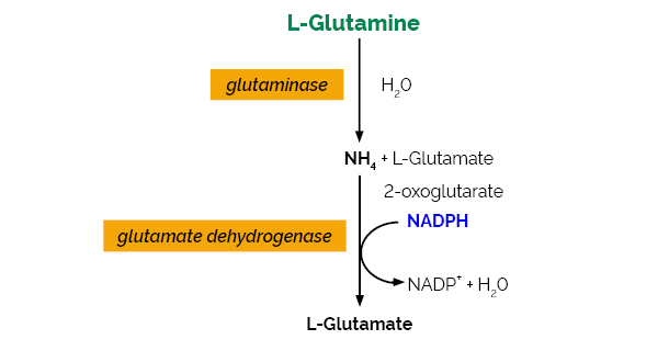
L-Glutamine/Ammonia Assay Kit (Rapid)
50 assays of each (manual) / 500 assays of each (microplate)
This product has been discontinued
Long term stability: See individual component labels
0.06 mg/L (ammonia)
Advantages
- Extended cofactors stability. Dissolved cofactors stable for > 1 year at 4oC.
- Very rapid reaction due to use of high activity glutaminase and uninhibited glutamate dehydrogense
- All enzymes supplied as stabilised suspensions
- Only enzymatic kit available
- Very cost effective
- All reagents stable for > 2 years after preparation
- Mega-Calc™ software tool is available from our website for hassle-free raw data processing
- Standard included
This product has been discontinued.
The L-Glutamine/Ammonia (Rapid) test kit is a novel method for the specific, convenient, cost effective and rapid measurement and analysis of L-glutamine and ammonia in culture media/supernatants and other materials.
Note for Content: The number of manual tests per kit can be doubled if all volumes are halved. This can be readily accommodated using the MegaQuantTM Wave Spectrophotometer (D-MQWAVE).
See our full list of assay kits.
Related Products
Similar Products
Related Products
Publications
New insights in the targets of action of dimethyl fumarate in endothelial cells: effects on energetic metabolism and serine synthesis.
Ocaña, M. C., Yang, C., Bernal, M., Martínez-Poveda, B., Vu, H., Cárdenas, C., DeBerardinis, R. & Medina, M. Á. Europe PMC, (2022), PPR490973.
New insights in the targets of action of dimethyl fumarate in endothelial cells: effects on energetic metabolism and serine synthesis.
Ocaña, M. C., Yang, C., Bernal, M., Martínez-Poveda, B., Vu, H., Cárdenas, C., DeBerardinis, R. & Medina, M. Á. Europe PMC, (2022), PPR490973.
Dimethyl fumarate is an ester from the Krebs cycle intermediate fumarate. This drug is approved and currently used for the treatment of psoriasis and multiple sclerosis, and its anti-angiogenic activity was reported some years ago. Due to the current clinical relevance of this compound and the recently manifested importance of endothelial cell metabolism on the angiogenic switch, we wanted to elucidate whether dimethyl fumarate has an effect on energetic metabolism of endothelial cells. Different experimental approximations were performed in endothelial cells, including proteomics, isotope tracing and metabolomics experimental approaches, in this work we studied the possible role of dimethyl fumarate in endothelial cell energetic metabolism. We demonstrate for the first time that dimethyl fumarate promotes glycolysis and diminishes cell respiration in endothelial cells, which could be a consequence of a down-regulation of serine and glycine synthesis through inhibition of PHGDH activity in these cells. Dimethyl fumarate alters the energetic metabolism of endothelial cells through an unknown mechanism, which could be the cause or the consequence of its pharmacological activity. This new discovery on the targets of this compound could open a new field of study regarding the mechanism of action of dimethyl fumarate.
Metabolic and Transcriptional Changes across Osteogenic Differentiation of Mesenchymal Stromal Cells.
Sigmarsdottir, T. B., McGarrity, S., de Lomana, A. L. G., Kotronoulas, A., Sigurdsson, S., Yurkovich, J. T., Rolfsson, O. & Sigurjonsson, O. E. (2021). Bioengineering, 8(12), 208.
Metabolic and Transcriptional Changes across Osteogenic Differentiation of Mesenchymal Stromal Cells.
Sigmarsdottir, T. B., McGarrity, S., de Lomana, A. L. G., Kotronoulas, A., Sigurdsson, S., Yurkovich, J. T., Rolfsson, O. & Sigurjonsson, O. E. (2021). Bioengineering, 8(12), 208.
Mesenchymal stromal cells (MSCs) are multipotent post-natal stem cells with applications in tissue engineering and regenerative medicine. MSCs can differentiate into osteoblasts, chondrocytes, or adipocytes, with functional differences in cells during osteogenesis accompanied by metabolic changes. The temporal dynamics of these metabolic shifts have not yet been fully characterized and are suspected to be important for therapeutic applications such as osteogenesis optimization. Here, our goal was to characterize the metabolic shifts that occur during osteogenesis. We profiled five key extracellular metabolites longitudinally (glucose, lactate, glutamine, glutamate, and ammonia) from MSCs from four donors to classify osteogenic differentiation into three metabolic stages, defined by changes in the uptake and secretion rates of the metabolites in cell culture media. We used a combination of untargeted metabolomic analysis, targeted analysis of 13C-glucose labelled intracellular data, and RNA-sequencing data to reconstruct a gene regulatory network and further characterize cellular metabolism. The metabolic stages identified in this proof-of-concept study provide a framework for more detailed investigations aimed at identifying biomarkers of osteogenic differentiation and small molecule interventions to optimize MSC differentiation for clinical applications.
NH4+ Suppresses NO3- Dependent Lateral Root Growth and Alters Gene Expression and Gravity Response in OsAMT1 RNAi Mutants of Rice (Oryza sativa).
Kumar, V., Kim, S. H., Priatama, R. A., Jeong, J. H., Adnan, M. R., Saputra, B. A., Kim, C. M., Je, B. II.,Park, S. J., Jung, K. H., Kim, K. M., Xuan, Y. H. & Han, C. D. (2020). Journal of Plant Biology, 63(5), 391-407.
The AMT1 family comprises major ammonium transporters in rice roots. In this study, we utilized AMT1 RNAi mutants (amt1) to explore how AMT1 affects NH4+- and NO3–-mediated morphological development and NH4+-responsive gene expression in roots. In the presence of NH4+, amt1 showed inhibition of NO3–- dependent lateral root development. The inhibitory action of NH4+ on lateral root growth was independent of the NO3– concentrations supplied to amt1 roots. The results of split root assays indicated that NH4+ exerts systemic action in inhibiting NO3–-dependent lateral root development in amt1. Further study with NAA and NOA, a potent auxin flux inhibitor, suggested that perturbation of membrane dynamics might not be the primary cause of the inhibitory action of NH4+ on NO3–-mediated lateral root growth in amt1 mutants. RNA-seq analysis of NH4+-responsive genes showed that approximately half of DEGs observed in wild-type roots were not detected in the DEGs of amt1 roots. Gene ontology enrichment analysis suggested that the expression of specific functional gene groups were affected by amt1 during the early response to NH4+. Auxin-responsive gene expression and root gravity responses were altered in amt1. This study demonstrated that AMT1 affects the interactions not only between ammonium and nitrate in lateral root growth but also between auxin and NH4+ in rice roots.
LC-MS/MS-based quantitative proteomic and phosphoproteomic analysis of CHO-K1 cells adapted to growth in glutamine-free media.
Kaushik, P., Curell, R. V. B., Henry, M., Barron, N. & Meleady, P. (2020). Biotechnology Letters, 42(12), 2523-2536.
LC-MS/MS-based quantitative proteomic and phosphoproteomic analysis of CHO-K1 cells adapted to growth in glutamine-free media.
Kaushik, P., Curell, R. V. B., Henry, M., Barron, N. & Meleady, P. (2020). Biotechnology Letters, 42(12), 2523-2536.
Objectives: This study aims to provide insights into the molecular mechanisms underlying adaptation of CHO-K1 cells to growth in glutamine-free media and potentially identifying critical signalling proteins and pathways involved in this phenotype. Results: A CHO-K1 cell line adapted to growth in glutamine-free media was established using a straightforward one-step glutamine reduction strategy. The adapted cell line had a comparable phenotype to the parental cells in terms of cell growth and viability. Global quantitative proteomic and phosphoproteomic analysis was carried out to compare the cells adapted to growth in glutamine-free media to parental cells grown in media containing 8 mM L-glutamine. The adaptation process was accompanied by changes in proteins associated with cytoskeleton rearrangement and mRNA splicing as evidenced via functional analysis of 194 differentially expressed proteins between the two cell lines. 434 phosphoproteins with altered abundance were also identified as a result of adaptation to L-glutamine-free conditions with an associated enrichment of pathways associated with MAPK and calcium signalling. Conclusions: This work provides a comprehensive proteomic and phosphoproteomic analysis of protein expression changes after adaptation to glutamine-free growth conditions highlighting critical pathways to consider in the rational design of improved feeding strategies or in cell line engineering to improve bioprocess phenotypes.
Development of a method for live octopus (Octopus vulgaris) transportation for long distance at high densities.
Araújo, J., Matias, A. C., Pousão‐Ferreira, P. & Soares, F. (2020). Aquaculture Research, 51(9), 3751-3759.
Development of a method for live octopus (Octopus vulgaris) transportation for long distance at high densities.
Araújo, J., Matias, A. C., Pousão‐Ferreira, P. & Soares, F. (2020). Aquaculture Research, 51(9), 3751-3759.
The development of new octopus‐based products with a growing economic value ensures the commercial interest of this species, making live octopus export an activity of great interest. The aim of this study was to develop a method for long‐distance transportation of live octopus at high densities. The system was composed by 220‐L tanks with cooling and aeration, where the animals were kept separated from each others. The water temperature was maintained at 10°C, after a decreasing of 1°C/hr. Live octopus transportation was tested for 48 hr at two densities: 50 kg/m3 and 100 kg/m3. During this period, water parameters were monitored. Stress response was evaluated through the analysis of haemolymph, muscle and brain tissues. No mortality was registered after 48 hr for both treatments. In all trials, water quality remained within the normal limits in both densities; there was, however, a significative increase of ammonia levels in the water. Ammonia, dopamine and Hsp70 levels were analysed in the beginning and at the end of the experiment for both densities; however, no significant differences were found among them. In general, this system seems to be a viable solution for live octopus 48‐hr transportation at a density of 100 kg/m3.
Three-layered silk fibroin tubular scaffold for the repair and regeneration of small caliber blood vessels: from design to in vivo pilot tests.
Alessandrino, A., Chiarini, A., Biagiotti, M., Dal Prà, I., Bassani, G. A., Vincoli, V., Settembrini, P., Pierimarchi, P., Freddi, G. & Armato, U. (2019). Frontiers in Bioengineering and Biotechnology, 7, 356.
Silk fibroin (SF) is an eligible biomaterial for the development of small caliber vascular grafts for substitution, repair, and regeneration of blood vessels. This study presents the properties of a newly designed multi-layered SF tubular scaffold for vascular grafting (SilkGraf). The wall architecture consists of two electrospun layers (inner and outer) and an intermediate textile layer. The latter was designed to confer high mechanical performance and resistance on the device, while electrospun layers allow enhancing its biomimicry properties and host's tissues integration. In vitro cell interaction studies performed with adult Human Coronary Artery Endothelial Cells (HCAECs), Human Aortic Smooth Muscle Cells (HASMCs), and Human Aortic Adventitial Fibroblasts (HAAFs) demonstrated that the electrospun layers favor cell adhesion, survival, and growth. Once cultured in vitro on the SF scaffold the three cell types showed an active metabolism (consumption of glucose and glutamine, release of lactate), and proliferation for up to 20 days. HAAF cells grown on SF showed a significantly lower synthesis of type I procollagen than on polystyrene, meaning a lower fibrotic effect of the SF substrate. The cytokine and chemokine expression patterns were investigated to evaluate the cells' proliferative and pro-inflammatory attitude. Interestingly, no significant amounts of truly pro-inflammatory cytokines were secreted by any of the three cell types which exhibited a clearly proliferative profile. Good hemocompatibility was observed by complement activation, hemolysis, and hematology assays. Finally, the results of an in vivo preliminary pilot trial on minipig and sheep to assess the functional behavior of implanted SF-based vascular graft identified the sheep as the more apt animal model for next medium-to-long term preclinical trials.
Liver-type glutaminase GLS2 is a druggable metabolic node in luminal-subtype breast cancer.
Lukey, M. J., Cluntun, A. A., Katt, W. P., Miao-chong, J. L., Druso, J. E., Ramachandran, S., Erickson, J. W., Le, H. H., Wang, Z. E., Blank, B., Greene, K. S. & Cerione, R. A. (2019). Cell Reports, 29(1), 76-88.
Efforts to target glutamine metabolism for cancer therapy have focused on the glutaminase isozyme GLS. The importance of the other isozyme, GLS2, in cancer has remained unclear, and it has been described as a tumor suppressor in some contexts. Here, we report that GLS2 is upregulated and essential in luminal-subtype breast tumors, which account for >70% of breast cancer incidence. We show that GLS2 expression is elevated by GATA3 in luminal-subtype cells but suppressed by promoter methylation in basal-subtype cells. Although luminal breast cancers resist GLS-selective inhibitors, we find that they can be targeted with a dual-GLS/GLS2 inhibitor. These results establish a critical role for GLS2 in mammary tumorigenesis and advance our understanding of how to target glutamine metabolism in cancer.
Daud, H., Browne, S., Al-Majmaie, R., Murphy, W. & Al-Rubeai, M. (2016). New Biotechnology, 33(1), 179-186.
Daud, H., Browne, S., Al-Majmaie, R., Murphy, W. & Al-Rubeai, M. (2016). New Biotechnology, 33(1), 179-186.
An understanding of the metabolic profile of cell proliferation and differentiation should support the optimization of culture conditions for hematopoietic stem and progenitor cell (HSPC) proliferation, differentiation, and maturation into red blood cells. We have evaluated the key metabolic parameters during each phase of HSPC culture for red blood cell production in serum-supplemented (SS) and serum-free (SF) conditions. A simultaneous decrease in growth rate, total protein content, cell size, and the percentage of cells in the S/G2 phase of cell cycle, as well as an increase in the percentage of cells with a CD71-/GpA+ surface marker profile, indicates HSPC differentiation into red blood cells. Compared with proliferating HSPCs, differentiating HSPCs showed significantly lower glucose and glutamine consumption rates, lactate and ammonia production rates, and amino acid consumption and production rates in both SS and SF conditions. Furthermore, extracellular acidification was associated with late proliferation phase, suggesting a reduced cellular metabolic rate during the transition from proliferation to differentiation. Under both SS and SF conditions, cells demonstrated a high metabolic rate with a mixed metabolism of both glycolysis and oxidative phosphorylation (OXPHOS) in early and late proliferation, an increased dependence on OXPHOS activity during differentiation, and a shift to glycolytic metabolism only during maturation phase. These changes indicate that cell metabolism may have an important impact on the ability of HSPCs to proliferate and differentiate into red blood cells.
Barth, C., Gouzd, Z. A., Steele, H. P., & Imperio, R. M. (2010). Journal of Experimental Botany, 61(2), 379-394.
Barth, C., Gouzd, Z. A., Steele, H. P., & Imperio, R. M. (2010). Journal of Experimental Botany, 61(2), 379-394.
Ascorbic acid (AA) is an antioxidant fulfilling a multitude of cellular functions. Given its pivotal role in maintaining the rate of cell growth and division in the quiescent centre of the root, it was hypothesized that the AA-deficient Arabidopsis thaliana mutants vtc1-1, vtc2-1, vtc3-1, and vtc4-1 have altered root growth. To test this hypothesis, root development was studied in the wild type and vtc mutants grown on Murashige and Skoog medium. It was discovered, however, that only the vtc1-1 mutant has strongly retarded root growth, while the other vtc mutants exhibit a wild-type root phenotype. It is demonstrated that the short-root phenotype in vtc1-1 is independent of AA deficiency and oxidative stress. Instead, vtc1-1 is conditionally hypersensitive to ammonium (NH4+). To provide new insights into the mechanism of NH4+ sensitivity in vtc1-1, root development, NH4+ content, glutamine synthetase (GS) activity, glutamate dehydrogenase activity, and glutamine content were assessed in wild-type and vtc1-1 mutant plants grown in the presence and absence of high NH4+ and the GS inhibitor MSO. Since VTC1 encodes a GDP-mannose pyrophosphorylase, an enzyme generating GDP-mannose for AA biosynthesis and protein N-glycosylation, it was also tested whether protein N-glycosylation is affected in vtc1-1. Furthermore, since root development requires the action of a variety of hormones, it was investigated whether hormone homeostasis is linked to NH4+ sensitivity in vtc1-1. Our data suggest that NH4+ hypersensitivity in vtc1-1 is caused by disturbed N-glycosylation and that it is associated with auxin and ethylene homeostasis and/or nitric oxide signalling.
Safety Information
Download Safety Data Sheet
Select a specific region and the Safety Data Sheet will open in a new window. Please make sure your browser doesn’t block pop-up windows

Receive Megazyme’s latest articles, news, product introductions and promotions
Customers also viewed
")
60 assays (manual) / 600 assays (microplate) / 700 assays (auto-analyser)
")
100 assays (manual) / 1000 assays (microplate) / 1000 assays (auto-analyser)
")
100 assays (manual) / 1000 assays (microplate) / 1000 assays (auto-analyser)
 (R-BAMR3)")
")
60 assays (manual) / 600 assays (microplate) / 600 assays (auto-analyser)
")
50 assays (manual) / 500 assays (microplate) / 500 assays (auto-analyser)


